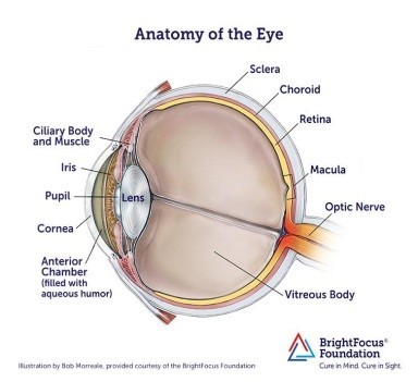
– Role of BCR in lymphoid tumor development: oculocerebral lymphoma (LOC) model (M Le Garff-Tavernier, C. Bravetti, F Davi). Primary central nervous system lymphoma (PCNSL) is a rare and severe form of diffuse large-cell B lymphoma, confined to the brain, spinal cord and meninges, often with ocular involvement but without peripheral extension. Pitié-Salpêtrière Hospital has an exceptional recruitment record for this type of tumor and is a national expert center for the coordination of the LOC network accredited by INCa. Our team has been working for years to optimize biological diagnosis and patient follow-up.
Our laboratory has developed a wide range of cellular techniques (cytology, multiparametric flow cytometry, lymphoid clonality testing, and targeted mutation testing by NGS) to optimize the detection of lymphoma cells in valuable fluids such as cerebrospinal fluid (CSF) and intraocular samples (aqueous humor and vitreous) (Armand Met al, Am J Hematol. 2019 and Bravetti C et al, Br J Haematol. 2023). We have recently started a collaboration with Dr. B. Mathon’s team, who provide us with irrigation fluid from stereotactic biopsies for diagnostic purposes. This cell-rich fluid allows us to provide rapid results within a day using cytology/cytometry techniques that complement the histology performed directly on the biopsy, thus optimizing the patient’s diagnostic pathway. We have also developed complementary approaches to quantify soluble biomarkers such as IL-10 and IL-6 in intraocular fluids. IL-10 also shows great promise as a marker for disease monitoring (response to treatment, disease progression or relapse), and our recent work indicates that it could be used as a marker of minimal residual disease (CAR-T cell therapy). We will continue this work by evaluating other soluble biomarkers, such as the identified prognostic indicators sCD19, CXCL13 and sCD25. We have been a reference medical biology laboratory (LBMR) for the diagnosis of oculocerebral lymphomas and we are an expert center in the LOC national network, a national medical expert network created in 2011 as part of a Cancer Plan, recognized and financed by the French National Cancer Institute. In this context, we participate in the network’s protocols, in particular PRT-K LOC-R01 for cytokine assays.
In a previous work, we demonstrated in primary vitreoretinal lymphoma (PVRL, a variety of LOC), and to a lesser extent in PCNSL, a major repertoire bias of the Ig variable regions of their BCR, with in addition an intraclonal diversity linked to the SHM process (Belhouachi N et al, Blood Adv. 2020). These data suggest the existence of continuous antigenic stimulation of tumor cells in vivo, which could play an important role in tumor growth. However, these results need to be refined with more resolutive techniques (NGS). We then plan to characterize their BCR repertoire in detail by NGS sequencing of the variable regions of their Ig heavy and light chains. In particular, we will investigate the clonal architecture of their BCR using the ViCloD software mentioned above. This will be compared with the clonal architecture at the oncogenic level. This work will be complemented by single cell analyses.
We also postulate that tumor cells can recognize antigens in situ that favor their ectopic localization and development. We have established a collaboration with Dr. K. Stomatopoulos (CERTH, Thessaloniki, Greece) whose team has the expertise to produce recombinant antibodies from BCRs derived from leukemia cells. Recombinant antibodies from PVRL BCR are currently being synthesized. They will be tested on “protein arrays” (self-antigens, microbial antigens), protein extracts from eye and brain biopsies, and on sections of these same tissues. The most relevant target antigens will be functionally tested on B lymphoma lines transfected with the corresponding BCRs.


