

Imaging
“A picture is worth 1000 words”
Confusius
“A picture is worth 1000 datas”
the CHICS
PRESENTATION
The imaging facility offers state-of-the-art infrastructure and expertise to meet your microscopy needs. We offer a range of microscopes from conventional to confocal microscopy.
We offer slide scanning services, training in the autonomous use of equipment and image analysis. We also foster close collaborations for in-depth analysis or the development of artificial intelligence analysis.
services
The imaging department offers :
- Advice to optimize the image quality of your markings.
- Assistance and training in instrument use.
- Slide scanning services.
- Image analysis and training.
- Collaborative work.
equipments
Zeiss – AxioObserver.Z1
Wide Field microscope: bright field, polarized light and fluorescence
Fluorescence: 4 parameters
DAPI*(Ex=365nm; Em=445nm)
FITC* (Ex=470nm; Em=525nm)
TRITC* (Ex=545nm; Em=605nm)
Cy5* (Ex=640nm; Em=690nm)
*or Compatible Fluorochromes

Leica Stellaris 5
Confocal Laser scanning microscope
3D imaging of thick specimens up to100µm
Excitations : 405nm, 448nm, white laser from 485nm to780 nm
5 detectors with adjustable spectral detection bands.
Easy imaging of 5 fluorochromes, (>10 possible).
Exemple of compatible fluorochromes :
- DAPI + Alexa 488, 594, 647, 750
- DAPI,+ Opal 520, 620, 690, 780
- Almost any fluorochrome can be used, however combinaisons of fluorochromes have to be choosen carefuly.

Scanner Zeiss – AxioScan.Z1
Slide scanner – Capacity 100 slides – Bright field and fluorescence
Fluorescence: 5 parameters
DAPI* (Ex=365nm; Em=445nm)
FITC* (Ex=470nm; Em=525nm)
TRITC* (Ex=545nm; Em=605nm)
Cy5* (Ex=640nm; Em=690nm)
Cy7* (Ex=735nm; Em=809nm)
*or Compatible Fluorochromes
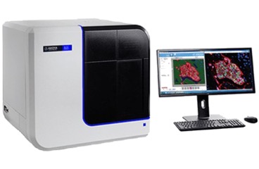
Scanner spectral Akoya – PhenoImager HT
80 slides capacity – Bright field and fluorescence
Demixing and tissue autofluorescence substraction
Fluorescence: 7 parameters
DAPI* (Ex=391nm; Em=453nm)
Opal480* (Ex=524nm; Em= 478nm)
Opal520* (Ex=486nm; Em=532nm)
Opal570* (Ex=538nm; Em=571nm)
Opal620* (Ex=590nm; Em=633nm)
Opal690* (Ex=699nm; Em=709nm)
Opal780* (Ex=735nm; Em=832nm)
*or Compatible Fluorochromes
SOFTWARES

Advanced image analysis software (qualitative, quantitative, supervised and unsupervised, cell segmentation, etc.) offering automation for data quantification.
Available on a computer workstation at CHICS – Free training
ZEN is the universal user interface used by all ZEISS imaging systems to capture and process images.
QuPath is an open-source image analysis software for the detection, segmentation and quantification of cells and tissues in histological images.
Free training
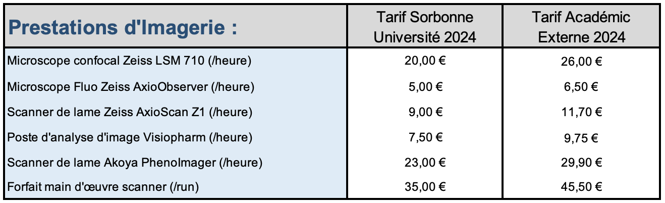
FEES
CONTACT
Christophe KLEIN
Email : christophe.klein@crc.jussieu.fr