20/12/2024
The CRC invites you to discover its research through beautiful scientific images from its labs. This is the “CRC Studio” series, to be followed throughout the year.
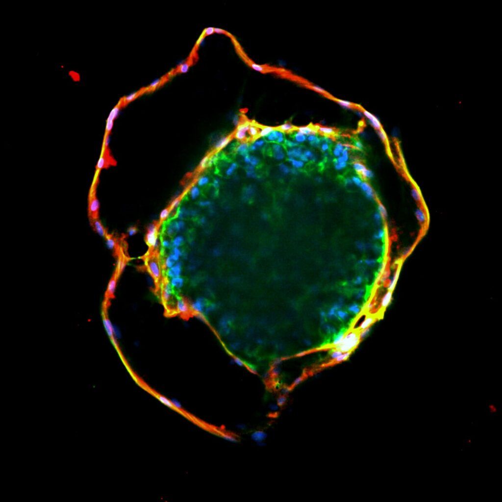
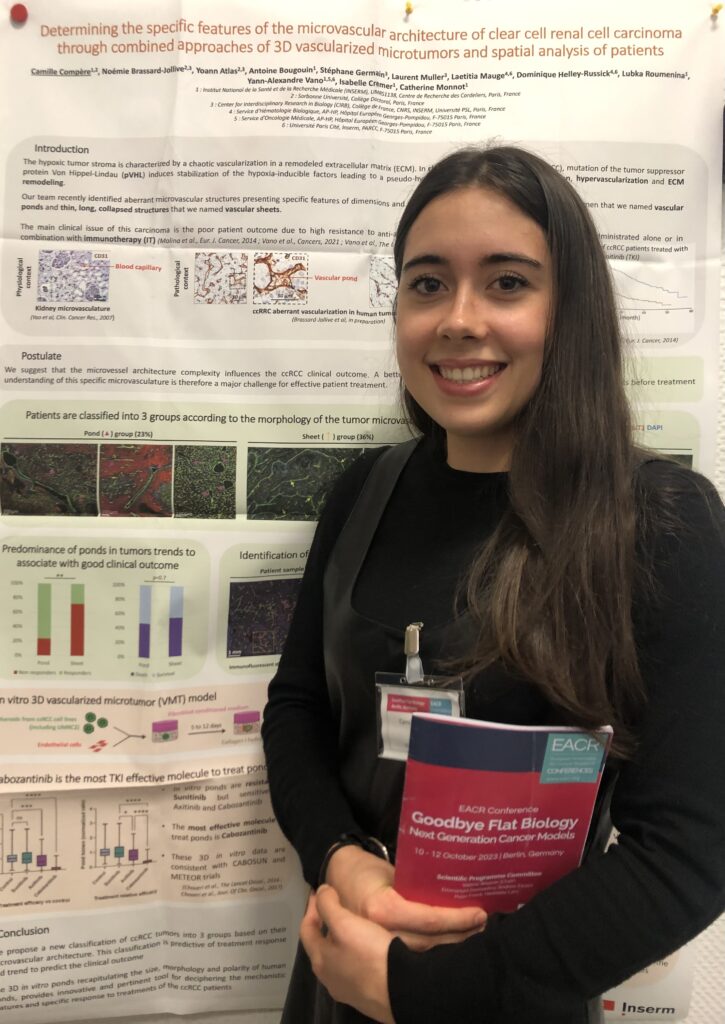
This image of a renal cell carcinoma microtumor “manufactured” in vitro in the laboratory was produced by Camille Compère, a doctoral student under the supervision of Catherine Monnot, in the Inflammation, Complement and Cancer team, headed by Isabelle Cremer.
The tumor mass, in the center, is made up of densely aggregated cancer cells (nucleus in blue, cytoplasm in green) surrounded by a blood vessel (in red) whose cavity (in black) is highly dilated and irregularly shaped. This microtumor grows within a collagen gel, enabling three-dimensional culture.

Clear cell renal cell carcinoma (ccRCC) microtumor produced in vitro in the laboratory.
Of particular interest to Camille are the architectural similarities between the blood vessels of patient tumors and those of microtumors fabricated in the laboratory, both of which present aberrant shapes and sizes, up to 100 times greater than a normal blood vessel.
Why is Camille interested in blood vessels?

Clear cell renal cell carcinoma (ccRCC) is a highly aggressive, highly vascularized cancer. The blood vessels within these tumors are often abnormally dilated, and thus able to deliver considerable amounts of oxygen and nutrients to the tumor core, greatly promoting its growth within the kidney as well as the development of metastases throughout the body.
The first line of treatment for these cancers targets and alters blood vessels to block tumor growth. However, ccRCC tumors can develop resistance to these treatments, limiting their effectiveness. It is therefore essential to be able to study these tumors and their sensitivity to treatments.
Camille is developing an experimental approach that makes it possible to manufacture ccRCC microtumors in vitro, very close anatomically and functionally to the patient’s tumor. These microtumors are subjected to treatments similar to those delivered to patients to study response or resistance to current treatments and test new therapeutic targets.
Ultimately, the researchers hope to gain a better understanding of the role of blood vessels in clear cell renal cell carcinoma, and to be able to propose new therapeutic avenues for patients.
Using a confocal microscope, Camille was able to scan the volume of the microtumor from top to bottom, in 3D.

